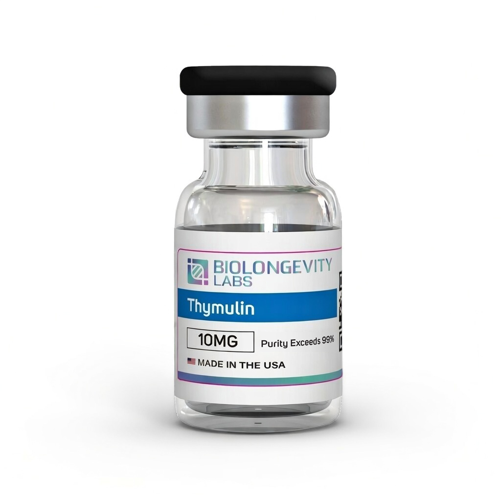
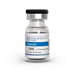
Thymulin Product Description
Thymulin belongs to the class of thymic peptides, a group of short signaling molecules originally isolated from thymic epithelial tissue. It was first described as facteur thymique sérique (FTS) and is also indexed as serum thymic factor and nonathymulin.
The molecule is a metallopeptide rather than a free peptide. Zinc coordination produces a defined three-dimensional conformation, and the zinc-free form does not carry the same recognition profile in binding assays [1].
Research interest sits across three areas: intrathymic and extrathymic T-cell differentiation, cytokine and NF-κB signaling in cultured cells and rodent models, and the interaction between thymic peptide output and neuroendocrine signaling [2].
Compound Specifications
| Property |
Value |
| CAS Number |
63958-90-7 |
| PubChem CID |
3085284 |
| Molecular Formula |
C₃₃H₅₄N₁₂O₁₅ |
| Molecular Weight |
858.86 g/mol |
| Amino Acid Sequence (three-letter) |
Pyr-Ala-Lys-Ser-Gln-Gly-Gly-Ser-Asn-OH |
| Amino Acid Sequence (one-letter) |
(pGlu)-AKSQGGSN |
| Sequence Length |
9 residues |
| Source |
Synthetic |
| InChIKey |
LIFNDDBLJFPEAN-BPSSIEEOSA-N |
| UNII |
9H198D04WL |
| Purity |
≥99% (HPLC) |
| Appearance |
White to off-white lyophilized powder |
| Solubility |
Soluble in sterile or bacteriostatic water |
| Storage |
-20°C, sealed, protect from light |
Storage and Handling
- Store the lyophilized compound sealed at -20°C, dry and protected from light.
- After reconstitution, store at 2°C to 8°C and use promptly.
- Avoid repeated freeze-thaw cycles, which reduce peptide integrity over successive handling.
- Maintain aseptic technique throughout reconstitution and aliquoting.
Lyophilized Format
This compound ships in lyophilized (freeze-dried) form. Freeze-drying supports long-term storage stability and preserves compound integrity. No fillers are added.
Thymulin Research Areas
The defining feature of thymulin in the published literature is zinc dependence. Nuclear magnetic resonance work established that zinc binding in an equimolar ratio produces a specific conformation, and that thymic epithelial cells release the peptide in its zinc-bound form [1].
In zinc-restricted animal models, measured thymulin activity falls in parallel with shifts in T-cell subpopulations and lymphokine output, and zinc repletion restores measured activity in the same systems [1]. Early characterization work described the molecule as a zinc-dependent metallopeptide acting on T-cell differentiation [3].
Signaling studies in rodent models have centered on the NF-κB pathway. In mice with induced neuroinflammation, thymulin administered alongside an IKK inhibitor reduced circulating interferon-γ and interleukin-6 and lowered phosphorylation of RelA and expression of heat shock protein Hsp72 in splenic lymphocytes [4]. A later study using thymulin bound to polybutylcyanoacrylate nanoparticles reported site-specific phosphorylation changes at RelA/p65 Ser276 and Ser536, along with reduced SAPK/JNK cascade activation [5].
Comparable mechanistic threads run through the wider bioregulator literature, summarized in our thymulin peptide research overview.
Beyond zinc binding and cytokine signaling, three further research threads appear consistently in the indexed literature.
Cytokine and Mediator Profiling in Rodent Models
A synthetic analogue of thymulin (PAT) was evaluated in endotoxin-challenged rats, where pretreatment lowered measured concentrations of interleukin-1β, interleukin-6, tumor necrosis factor-α, and nerve growth factor at the injection site, and reduced prostaglandin E₂ in liver tissue after systemic challenge [6].
In an ovalbumin-challenged airway model in mice, delivery of a thymulin analogue gene using compacted DNA nanoparticles reduced collagen deposition and smooth muscle hypertrophy in lung tissue, with plasmid detectable in lung tissue for up to 27 days [7].
Extrathymic Release Under Cellular Stress
Thymulin is not exclusively thymic in origin. When RAW 264.7 macrophages and L929 fibroblasts were exposed to oxidative stress, heat, apoptotic, or necrotic signals, extracellular thymulin appeared in the culture medium within two hours [8].
Western blot analysis of macrophage lysates identified bands at approximately 60 kDa and 10 kDa, interpreted as a candidate precursor and intermediate. A BLAST search against the thymulin sequence returned SPATS2L, an intranucleolar stress-response protein of about 62 kDa carrying a thymulin-like sequence [8].
Neuroendocrine Signaling
Review work characterizes thymulin as a hypophysiotropic peptide, with reported activity on pituitary hormone release in dispersed rat pituitary cell preparations, and with its own output influenced in turn by growth hormone, prolactin, and thyroid hormone signaling [2].
The same review notes that an adenoviral vector carrying a synthetic thymulin gene achieved longer transgene expression in rat brain than adenovirally mediated expression of other reporter genes, with local cytokine modulation proposed as the mechanism [2].
| Research Area |
In Vitro Application |
| Metallopeptide chemistry |
Zinc coordination studies and conformational analysis by NMR |
| Immune cell signaling |
T-cell differentiation assays in cultured lymphocyte lines |
| Cytokine pathway research |
NF-κB and SAPK/JNK cascade profiling in stimulated cell cultures |
| Cell stress response |
Detection of extracellular peptide release in stressed macrophage and fibroblast cultures |
| Analytical method development |
HPLC and mass spectrometry reference standard for nonapeptide characterization |
References
- Dardenne M, Pleau JM (1994). Interactions between zinc and thymulin. Metal-Based Drugs. https://doi.org/10.1155/MBD.1994.233
- Reggiani PC, Morel GR, Cónsole GM, et al. (2009). The thymus-neuroendocrine axis: physiology, molecular biology, and therapeutic potential of the thymic peptide thymulin. Annals of the New York Academy of Sciences. https://doi.org/10.1111/j.1749-6632.2008.03964.x
- Bach JF, Dardenne M (1989). Thymulin, a zinc-dependent hormone. Medical Oncology and Tumor Pharmacotherapy. https://doi.org/10.1007/BF02985220
- Lunin SM, Khrenov MO, Novoselova TV, et al. (2015). Modulation of inflammatory response in mice with severe autoimmune disease by thymic peptide thymulin and an inhibitor of NF-kappaB signalling. International Immunopharmacology. https://doi.org/10.1016/j.intimp.2015.01.021
- Lunin SM, Khrenov MO, Glushkova OV, et al. (2019). Protective Effect of PBCA Nanoparticles Loaded with Thymulin Against the Relapsing-Remitting Form of Experimental Autoimmune Encephalomyelitis in Mice. International Journal of Molecular Sciences. https://doi.org/10.3390/ijms20215374
- Safieh-Garabedian B, Dardenne M, Pléau JM, Saadé NE (2002). Potent analgesic and anti-inflammatory actions of a novel thymulin-related peptide in the rat. British Journal of Pharmacology. https://doi.org/10.1038/sj.bjp.0704793
- da Silva AL, Martini SV, Abreu SC, et al. (2014). DNA nanoparticle-mediated thymulin gene therapy prevents airway remodeling in experimental allergic asthma. Journal of Controlled Release. https://doi.org/10.1016/j.jconrel.2014.02.010
- Lunin SM, Khrenov MO, Glushkova OV, et al. (2017). Extrathymic production of thymulin induced by oxidative stress, heat shock, apoptosis, or necrosis. International Journal of Immunopathology and Pharmacology. https://doi.org/10.1177/0394632017694625
Certificate of Analysis (COA) for Every Batch
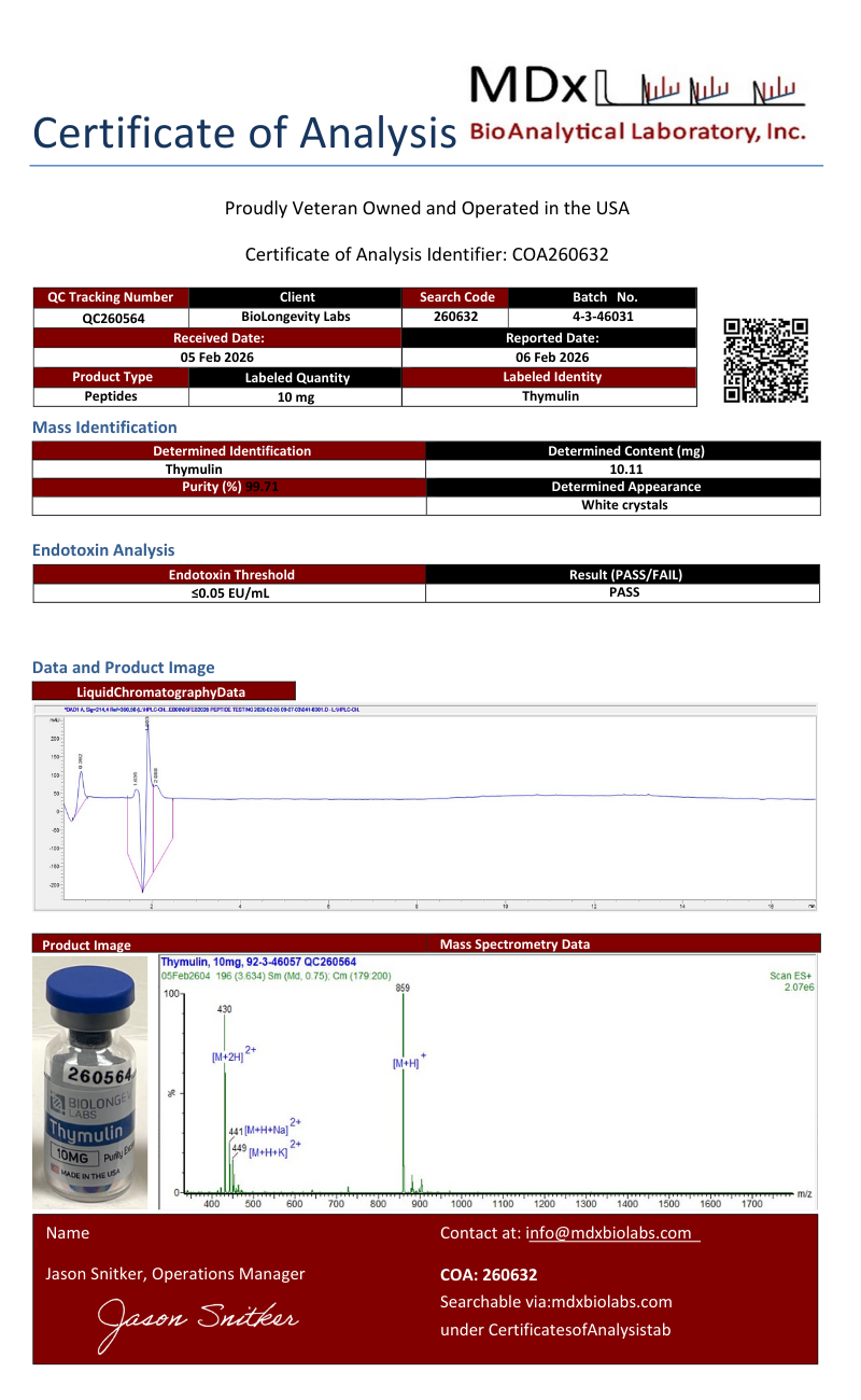
A Certificate of Analysis (COA) is a document that verifies a compound’s identity, purity, and batch quality through independent laboratory testing. Every compound from BioLongevity Labs ships with a COA tied to its specific batch, so researchers can confirm exactly what they received before it enters a protocol.
Each COA reports results from third-party laboratory analysis, including:
- Ultra-high-performance liquid chromatography with mass spectrometry (UHPLC-MS) for purity, typically confirmed at 99% or higher
- Mass identification for molecular confirmation and content quantitation
- Endotoxin quantitation by Limulus amebocyte lysate (LAL) assay where applicable
- Visual and physical characterization of the finished material
How to verify a COA independently
Every certificate can be checked against the issuing laboratory’s own records, not just the copy hosted here. Verification does not depend on BioLongevity Labs.
- MDx BioAnalytical Laboratory certificates carry a QC tracking number and a search code. Newer certificates also carry a QR code. Scan the code, or enter the search code at mdxbiolabs.com, to pull the official record.
- BioRegen reports of analysis carry a Report ID and a Validation Key. Scan the QR code on the certificate to open the official record, or reference both identifiers when contacting the laboratory at the address printed on the report.
- SafeCert Labs certificates, which appear on a number of earlier batches, carry a COA number and the signature of the reporting chemist. Reference that number when requesting confirmation from the laboratory directly.
Batches are frequently tested by both laboratories independently. When two certificates exist for the same lot, each one resolves at its own issuing laboratory, which lets a researcher confirm the same material twice through two unrelated sources.
COAs are sourced from independent certified labs rather than in-house testing alone, giving researchers a verifiable record of molecular integrity for each batch. All compounds are supplied for research use only.
Review the COAs for this batch below, or browse the full COA library.
Thymulin (260632)
×
![]()